Seeing the Full Picture: How Helical CT Scanning Transforms Inspection of Medical Implant Systems
Seeing the Full Picture: How Helical CT Scanning Transforms Inspection of Medical Implant Systems
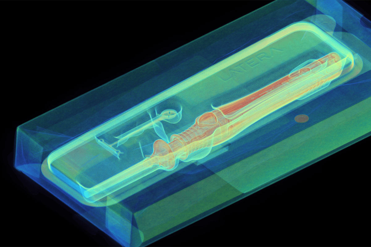
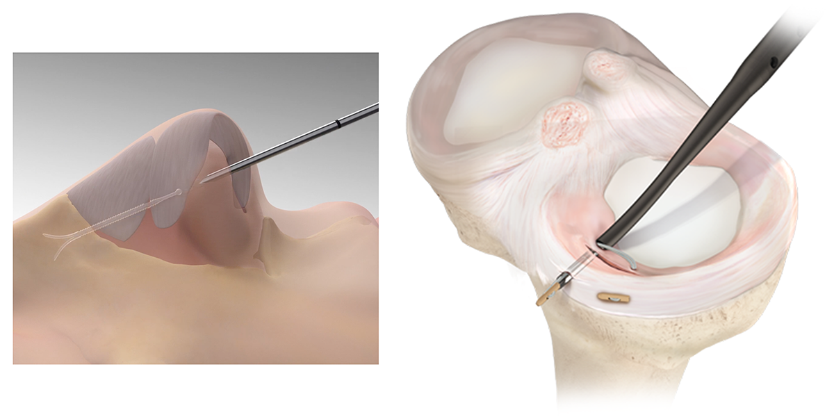
Medical implants are getting smaller, smarter, and more sophisticated. Their delivery instruments are getting longer and lower profile as the industry optimizes for minimally invasive procedures. For example, devices such as the LATERA® Nasal Implants and the Smith & Nephew OM-6500 SpeedScrew meniscus repair implants rely on elongated geometries to deliver strength, controlled deployment, and predictable performance inside the body. Their length is essential to how they function yet it also makes them notoriously difficult to inspect with traditional industrial CT scanning.
Conventional CT systems have a fixed vertical field of view, meaning long parts often can’t be captured in a single scan without compromising resolution. Engineers are forced into a tradeoff: pull the sample farther from the detector to fit the whole part and lose critical detail, or scan smaller sections and attempt to stitch the datasets together manually. Neither approach is ideal when you’re analyzing devices designed for precise deployment, controlled absorption, and long-term patient safety.
Why length matters for implant inspection
LATERA® nasal implants are designed to support nasal valve collapse, requiring thin, flexible long-form geometries that must be manufactured with extremely tight tolerances. Small deviations in wall thickness, anchor structure, or material uniformity can affect how the implant deploys or resorbs over time.
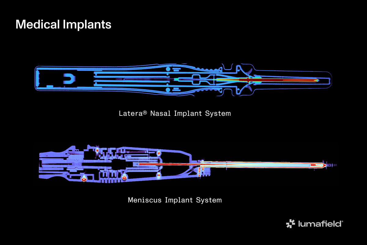
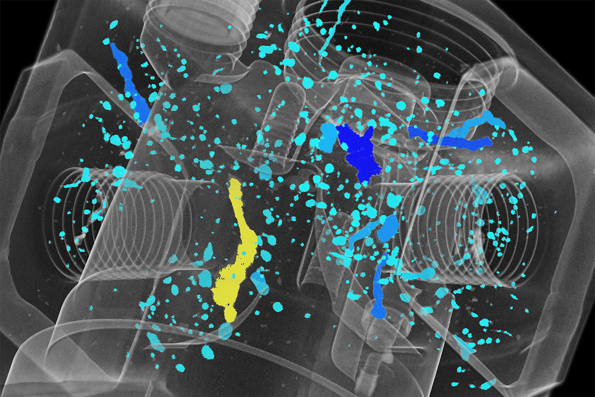
Meniscus repair implants (such as darts, anchors, and long fasteners) have even more complex requirements. Their strength, barbs or anchors, and delivery geometry span the full length of the device. Engineers often need to inspect barb geometry and sharpness, fiber reinforcement, internal alignment, or internal molding defects such as microcracks or voids along the length. But because these implants are long and slender, imaging the entire device at high resolution has traditionally been a challenge.
How Helical Scanning solves the problem
Helical Scanning removes the traditional vertical limitations of industrial CT. Instead of capturing a fixed-height volume, the system moves the part upward as it rotates, collecting a continuous helical dataset. This means for long medical implants, you no longer have to pull the implant farther from the detector to make it fit, or do traditional Industrial CT scan stitching that can introduce alignment errors. The system maintains optimal magnification while extending the scan vertically, making it ideal for slender devices like LATERA implants, suture anchors, and repair darts.
Seamless workflow, better results
Helical Scanning in Lumafield’s Voyager workflow is intentionally simple. Setting upper and lower bounds takes seconds, and the dataset opens like any other CT scan except with an extended field of view and dramatically more usable information. Every tool, measurement, and visualization works exactly as expected. And ultimately, this leads to safer, more reliable implantable products.