CT scanning Darwin’s finches at Cal Academy with Adam Savage
Museums are not just sites of preservation and teaching; they are also centers of cutting-edge research and technological innovation. The California Academy of Sciences in San Francisco welcomed Adam Savage and the Tested team for a behind-the-scenes visit to explore the creative ways curators are maintaining and reimagining their one-of-a-kind collections.
Founded over 150 years ago, Cal Academy houses both a world-class museum and the Institute for Biodiversity Science and Sustainability, home to over 46 million specimens from around the world. While a fraction of these specimens are on display for the public, the majority remain in storage and are used primarily for research purposes. These specimens, some dating back to the mid-1800s, offer a historical snapshot of biodiversity from about 174 countries.

CT and 3D digitization
Researchers are currently focused on digitizing their vast array of specimens, and modern scanning technologies—particularly CT scanning—allow Cal Academy to make their collection more accessible than ever before. The Academy is working on a project to digitize a significant portion of its Galapagos collections with 3D surface scanning and CT scanning. The goal is to scan one male and one female of every tetrapod (non-fish vertebrate) from every island where they are found. This ambitious project aims to scan about 1,300 specimens, with around 900 already completed.

Inside Darwin’s finches with CT
Cal Academy’s collection includes the same group of species that influenced Charles Darwin’s theory of natural selection, but it does not include the actual birds found on his voyages in the 1830s. Over time, these species of finches have collectively been dubbed “Darwin’s finches” in recognition of their historical and scientific significance, even though the specimens studied today are not directly from Darwin's original collection. Their diverse beak shapes and sizes, adapted to different food sources, provide a compelling example of adaptive radiation.

Jack Dumbacher, Curator of Ornithology and Mammalogy, sheds light on how modern technology, particularly CT scanning, is offering new insights into these iconic birds. Traditional methods of studying them involve physical examinations and measurements. With a CT scan, researchers can non-destructively visualize bone structures in great detail, allowing them to observe minute differences that might not be evident through external examination.
In the scan embedded below, color-coded density maps make denser areas like bones appear in red, while less dense areas, such as flesh and musculature, are shown in blue. This detailed view can help researchers understand the underlying skeletal structures that give rise to the diverse beak shapes.
While the primary focus might be on the beaks, CT scanning has also revealed unexpected findings. For instance, Dumbacher observed unusual patterns on the skin of some scanned birds. While the exact nature of these patterns remains a mystery, they could be indicative of conditions like Avian Pox. The CT scans can peer through the feathers, revealing potential lesions or abnormalities on the bird's skin that might otherwise go unnoticed.

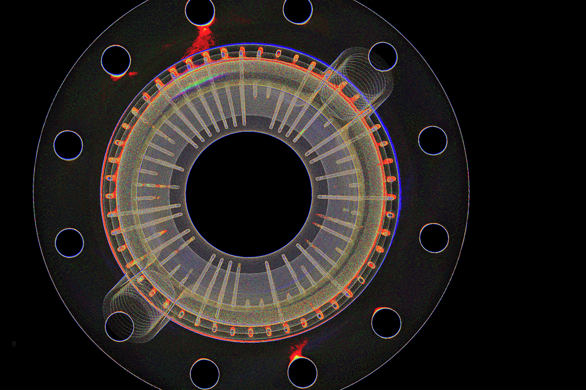
What looks like a coiled spring inserted into this finch’s neck is actually its trachea, or windpipe—the tube through which birds breathe. These coils are made up of cartilaginous rings, which can become bony as the bird matures. They prevent the trachea from collapsing when the bird inhales. Without them, the negative pressure created during inhalation would cause a simple tube to collapse, obstructing airflow.
Right next to the trachea, we note the presence of blue blobs, likely seeds the finch had consumed. Since they lack teeth, birds swallow seeds whole. These seeds are stored in a crop, a pouch-like structure, before moving down to the gizzard, a muscular part of the stomach that grinds the seeds.
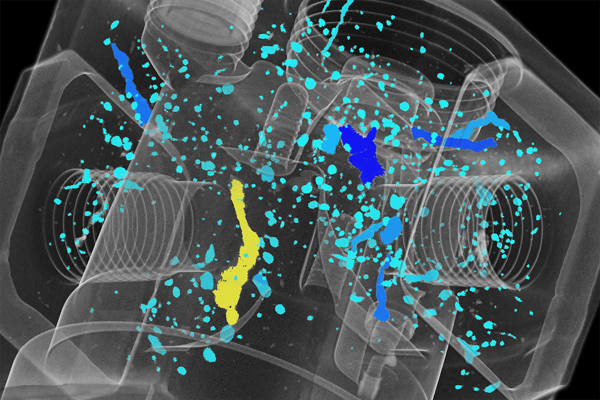
The ability to compare different specimens at a granular level is crucial for researchers. Using Voyager’s Scan to Scan Comparison feature, we compared two different scans of medium ground finch specimens to study the minute differences (see embed below). Precise comparisons like this one can provide insights into quantitative variations within the same species, potentially revealing evolutionary adaptations or anomalies.
Beyond research, the digitized scans of the finches have educational potential. By printing 3D models of the finch beaks, educators can offer students a tangible way to understand evolutionary concepts. These models can be used in practical demonstrations, allowing students to grasp the functional implications of the different beak shapes.
Anyone can explore these specimens from Cal Academy’s “library of life” for themselves right in their web browser with Lumafield's Voyager platform. Voyager is a cloud-based analysis software powered by AI, designed specifically for engineers and researchers to inspect, measure, and compare CT scans, CAD, STL, and 3D mesh files. Anyone can collaboratively access scan data from anywhere, without the need for specialized hardware.
Conclusion
The California Academy of Sciences carries the torch of its rich history of innovation by pursuing the most advanced technologies in the present. The insights gained from research not only enhance our understanding of animal biology but also underscore the importance of preserving specimens for future study. Their efforts in digitizing their collection, especially through CT scanning, ensure that the wealth of knowledge they hold is shared and preserved for generations to come.